KI-Beratung Beta

Gutscheinbedingungen

**Gültig bis 09.11.2026 auf tolino shine color (4016621130473), tolino vision color (4016621130183) und tolino vision color weiß (4016621131456) online auf thaalia.live, in der Thalia App und in allen Thalia Buchhandlungen in Österreich. In den Buchhandlungen nur gültig auf lagernde Ware. Pro Einkauf einmal einlösbar. Nur gültig gegen Vorlage oder im Onlineshop hinterlegter Bonuscard. Infos zur Einlösung in der Buchhandlung sind auf der Bonuscard-Vorteilspreisseite zu finden. Click & Collect nur bei Onlinevorabzahlung möglich. Keine Einlösung bei Scan & Go-Bezahlung. Keine Barauszahlung. Nicht kombinierbar mit anderen Aktionen und Gutscheinen. Nicht gültig für Geschenkkarten, Versandkosten und Services. Nur solange der Vorrat reicht.

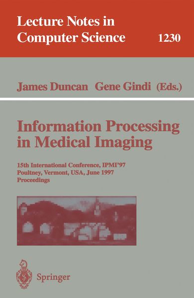
  • Produktbild: Information Processing in Medical Imaging
  • Produktbild: Information Processing in Medical Imaging
Band 1230

Information Processing in Medical Imaging 15th International Conference, IPMI'97, Poultney, Vermont, USA, June 9-13, 1997, Proceedings

51,99 €

inkl. gesetzl. MwSt., Versandkostenfrei


Beschreibung

Produktdetails

Einband

Taschenbuch

Erscheinungsdatum

21.05.1997

Abbildungen

XVI, 564 p.

Herausgeber

James Duncan + weitere

Verlag

Springer Berlin

Seitenzahl

564

Maße (L/B/H)

23,5/15,5/3,2 cm

Gewicht

867 g

Auflage

1997

Sprache

Englisch

ISBN

978-3-540-63046-3

Beschreibung

Produktdetails

Einband

Taschenbuch

Erscheinungsdatum

21.05.1997

Abbildungen

XVI, 564 p.

Herausgeber

Verlag

Springer Berlin

Seitenzahl

564

Maße (L/B/H)

23,5/15,5/3,2 cm

Gewicht

867 g

Auflage

1997

Sprache

Englisch

ISBN

978-3-540-63046-3

Herstelleradresse

Springer-Verlag KG
Sachsenplatz 4-6
1201 Wien
AT

Email: ProductSafety@springernature.com

Noch keine Bewertungen vorhanden

Verfassen Sie die erste Bewertung zu diesem Artikel

Helfen Sie anderen Kundinnen und Kunden durch Ihre Meinung.

Kundinnen und Kunden meinen

Bewertungen (0)

  • Produktbild: Information Processing in Medical Imaging
  • Produktbild: Information Processing in Medical Imaging
  • Automatic construction of eigenshape models by Genetic Algorithm.- Quadratic variation of deformations.- The softassign Procrustes matching algorithm.- Acoustic and elastic imaging to model disease-induced changes in soft tissue structure.- 2D intravascular E.I.T. using a non-iterative, non-linear reconstruction algorithm.- Optimal data types in optical tomography.- Using spatial-temporal images for analysis of gated cardiac tomographic data: The “M-Mode” revisited.- Automated segmentation of brain exterior in MR images driven by empirical procedures and anatomical knowledge.- Automated 3D segmentation using deformable models and fuzzy affinity.- Segmentation of medical image objects using deformable shape loci.- Mapping the cerebral sulci: Application to morphological analysis of the cortex and to non-rigid registration.- Bayesian detection of random signals on random backgrounds.- Approximate distributions for Maximum Likelihood and maximum a posteriori estimates under a Gaussian noise model.- Continuous Gaussian mixture modeling.- New statistical models for randoms-precorrected PET scans.- Registration of 3D medical images using simple morphological tools.- Deformation for image guided interventions using a three component tissue model.- Registration of abdominal CT and SPECT images using Compton scatter data.- Definition of volume transformations for volume interaction.- Nonlinear versus linear models in functional neuroimaging: Learning curves and generalization crossover.- Synthetic resampling methods for variance estimation in parametric images.- Space-time statistical model for functional MRI image sequences.- Jackknifing a normal database for regional cerebral blood flow SPECT obtained using principal components.- Generating 3-D cardiac material markersusing tagged MRI.- Identification of myocardial tags in tagged MR images without prior knowledge of myocardial contours.- An autofocus algorithm for the automatic correction of motion artifacts in MR images.- Measurement of global and regional cerebral volume changes by integrating boundary shifts between registered serial 3D MR scans.- Automatic detection of sulcal bottom lines in MR images of the human brain.- Segmentation and interpretation of MR brain images using an improved knowledge-based active shape model.- Gaussian random fields on sub-manifolds for characterizing brain surfaces.- Fuzzy logic approach to 3D magnetic resonance image segmentation.- Comparison of traditional brain segmentation tools with 3D self-organizing maps.- Finding the brain cortex using fuzzy segmentation, isosurfaces, and deformable surface models.- Statistical modelling of lines and structures in mammograms.- A thinning algorithm to extract medial lines from 3D medical images.- Setting the mind for intelligent interactive segmentation: Overview, requirements, and framework.- Segmentation of cerebral vessels and aneurysms from MR angiography data.- Computation of efficient patient specific models from 3-D medical images: Use in virtual endoscopy and surgery rehearsal.- In vivo analysis of trabecular bone architecture.- Automatic measurement of vertebral shape using Active Shape Models.- A hybrid approach for the detection of small airways disease from Computed Tomographic images.- Computer simulation of convection and diffusion effects on velocity estimations from X-ray contrast density time curves.- A comparison of retrospective intensity non-uniformity correction methods for MRI.- A robust and efficient algorithm for image registration.- Automatical adaption of the stereotactical coordinate system in brain MRI datasets.- Design of a statistical model of brain shape.- Automatic landmark identification using a new method of non-rigid correspondence.- Non-rigid curve correspondence for estimating heart motion.- Tracking of tagged MR images by Bayesian analysis of a network of quads.- Noninvasive skin imaging.- Modelling interreflections in a system based on the differential absorption of light (DAL) for the measurement of range images.- Quantitative estimation of scatterer spacing from backscattered ultrasound signals using the complex cepstrum.- Spatial compounding of 3-D ultrasound images.- Cone beam imaging of a section of a long object with a short detector.- New methods for SPECT imaging and pre-reconstruction restoration.- Reconstruction of the intracerebral vasculature from MRA and a pair of projection views.- Two alternative forced choice evaluation of vessel visibility increases due to zero-filled interpolation in MR angiography.- Bayesian detection with amplitude, scale, orientation and position uncertainty.